Disclaimer: This website provides health information for educational purposes only and is not a substitute for professional medical advice, diagnosis, or treatment. Always seek the guidance of a qualified healthcare provider with any questions you may have.
At many diagnostic centers today, one of the more common imaging procedures used nowadays is a PET (positron emission tomography} scan. This is among the latest technologies in nuclear medicine imaging. For some, the prospect of a PET scan may be uncomfortable. Today we’ll break down what actually happens during a PET, how it works, and how it can save your life.
The PET Scan Procedure
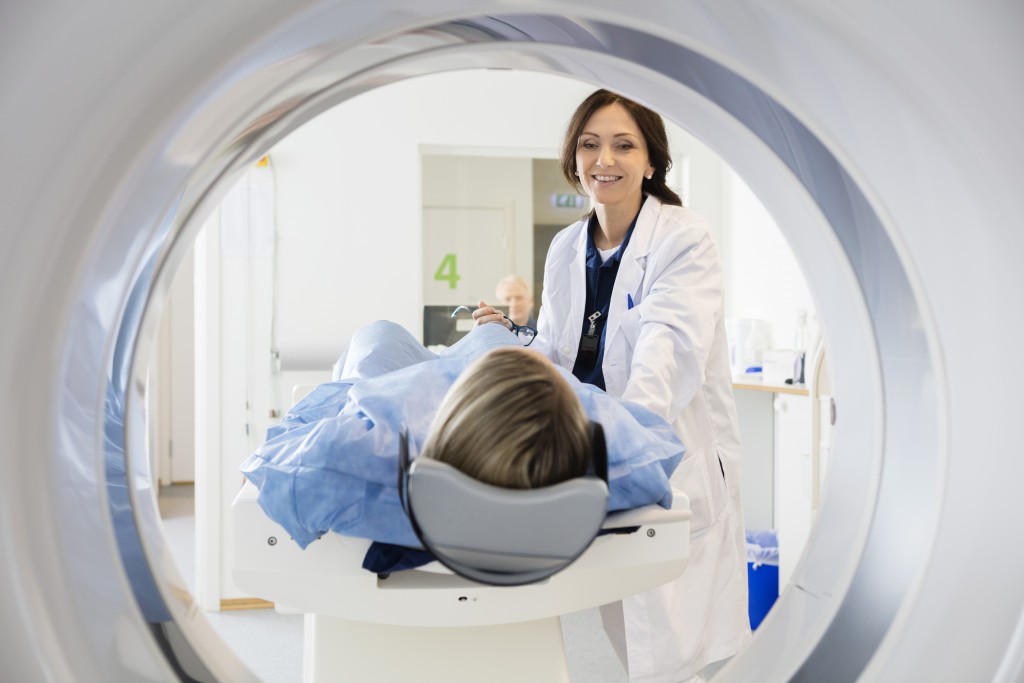
A radiolabelled molecule is be injected into the patient’s veins. Fluorodeoxyglucose is the common molecule used for PET scanning, usually given by injection. This sugar solution then accumulates in the body parts with high glycolic metabolism (such as tumors, which tend to use up more sugar in the body than normal tissue) and generates gamma rays. These rays are detected by a scanner that changes them into 3D images. The following are the typical conditions a doctor might want to pick when he/she orders a PET scan.
While the scan does expose the patient to small amounts of radiation, the benefits (accurate detection of cancer and other potentially life-threatening conditions) usually outweigh the risks. Doctors will also try to minimize the exposure by only scanning small areas.
What A PET Scan can Detect

- Cancer. Tumor cells have higher metabolic rates compared to other cells. This means a high glycolic activity that will be evidenced by bright spots in your PET scan. The scans in oncology are used for the diagnosis of cancer cells, staging of disease, and checking the effectiveness of treatment. It is also used to detect recurrence after you have finished a treatment course and been declared cancer-free. Even so, the scans are used in combination with other blood tests and imaging studies.
- Heart Conditions. Heart disease is the leading cause of death in most countries, according to the World Health Organization. Nowadays, PET scans are another tool in the physicians’ kit that allows the early diagnosis of heart issues and their optimal treatment. In the heart, the scans will pick the sections of decreased blood flow. Healthy heart tissue takes in more of the tracer compared to segments where there is reduced blood flow. The brightness and colors in different parts of the scan help doctors detect what areas of your heart have diseased tissue.
- Brain Disorders. The primary fuel in your brain is glucose. During a PET scan, the injected tracer will attach to different compounds. It will detect the sections where there is a high glucose activity. In these sections, you might need further tests to check the cause of the high glycolic activity. PET scans are used for the diagnosis of Parkinson’s, Alzheimer’s, depression, and epilepsy. The scan will also prove useful for assessing the extent of brain damage after head trauma
- Inflammatory Conditions.There is also a high glucose level in inflammatory diseases. As such, PET scans might also be used to detect inflammatory conditions. This is often in cases where blood markers of inflammation do not pinpoint the exact inflamed section.
People are apprehensive about PET scans because they hear about the risk of exposure to radiation. The cost, also, can be prohibitive, without a co-pay or insurer. In a privatediagnostic center in London, for example, a PET-CT can cost up to £2000. This cost, however is usually comparable to (or in some situations, more cost-efffective than) other diagnostic imaging tools like MRIs.
In the above instances, however, PET scans might be the best diagnostic tool for the early detection and treatment of many serious health issues. By carefully weighing a mitigated risk, PET scans are saving lives all over the world.


